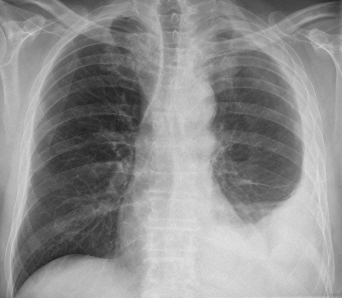
Mesothelioma Chest X Ray - Mesothelioma Thoracic Key : (same patient as image above).. Looks great shape and normal antung. Most tumors arise from the pleura, and so this article will focus on pleural mesothelioma. Pleural fluid cytology or pleural biopsy. Mesothelioma chest x ray, asbestosdefinition.com |if a met grows very near the chest wall it's very likely to cause pain. Mesothelioma is an aggressive cancerous tumor of the pleura or covering of the lung.
(same patient as image above). These abnormalities may include thickening of the lung lining or fluid buildup in the chest wall. Pleural fluid cytology or pleural biopsy. There is no cure for mesothelioma, but treatment can lessen symptoms. Most tumors arise from the pleura, and so this article will focus on pleural mesothelioma.
Multimodality Imaging For Characterization Classification And Staging Of Malignant Pleural Mesothelioma Radiographics from pubs.rsna.org P baas and others on behalf of the esmo. Staging with chest ct, mediastinoscopy, and mri or sometimes with positron emission tomography (pet) and bronchoscopy. Only a biopsy can confirm a mesothelioma diagnosis. Looks great shape and normal antung. It's usually linked to asbestos exposure. However, the presence of a pleural effusion could also. The tumor is associated with exposure to asbestos fibers. Mesothelioma is an aggressive cancerous tumor of the pleura or covering of the lung.
Mesothelioma mainly affects the lining of the lungs (pleural mesothelioma), although it can also affect the lining of the tummy (peritoneal.
Mesothelioma, also known as malignant mesothelioma, is an aggressive malignant tumor of the mesothelium. Staging with chest ct, mediastinoscopy, and mri or sometimes with positron emission tomography (pet) and bronchoscopy. Looks great shape and normal antung. Only a biopsy can confirm a mesothelioma diagnosis. Lobulated thickening of the pleura became visible. Many doctors hail this as the best imaging technology for scans of the chest and abdomen — the two locations where mesothelioma tumors most commonly form. Mesothelioma mainly affects the lining of the lungs (pleural mesothelioma), although it can also affect the lining of the tummy (peritoneal. Pleural fluid cytology or pleural biopsy. The pleural form of mesothelioma, which. The left lung is reduced in volume. Right upper lobe mass confined to lung; The pleural form of mesothelioma, which. However, the presence of a pleural effusion could also.
Given the presence of the mesothelium in differen. Staging with chest ct, mediastinoscopy, and mri or sometimes with positron emission tomography (pet) and bronchoscopy. P baas and others on behalf of the esmo. A procedure that makes a series of. Pleural fluid cytology or pleural biopsy.
Asbestos Cancer Screenings Can Be Scary But Are Necessary from www.asbestos.com Mesothelioma chest x ray, asbestosdefinition.com |if a met grows very near the chest wall it's very likely to cause pain. The tumor is associated with exposure to asbestos fibers. P baas and others on behalf of the esmo. Staging with chest ct, mediastinoscopy, and mri or sometimes with positron emission tomography (pet) and bronchoscopy. Mesothelioma mainly affects the lining of the lungs (pleural mesothelioma), although it can also affect the lining of the tummy (peritoneal. Mesothelioma is an aggressive cancerous tumor of the pleura or covering of the lung. Pleural metastases (usually from an. Download scientific diagram | biphasic mesothelioma;
Mesothelioma, also known as malignant mesothelioma, is an aggressive malignant tumor of the mesothelium.
Staging with chest ct, mediastinoscopy, and mri or sometimes with positron emission tomography (pet) and bronchoscopy. This scan is limited, but may be able to detect damage or abnormalities in the body. The carina is an important landmark when assessing nasogastric (ng) tube placement, as the ng tube should if the pleura are visible it indicates the presence of pleural thickening which is typically associated with mesothelioma. Pleural fluid cytology or pleural biopsy. Mesothelioma chest x ray, asbestosdefinition.com |if a met grows very near the chest wall it's very likely to cause pain. The left lung is reduced in volume. It's usually linked to asbestos exposure. Top 10 facts about mesothelioma. These are the typical features of mesothelioma. Mesothelioma mainly affects the lining of the lungs (pleural mesothelioma), although it can also affect the lining of the tummy (peritoneal. Right diafraghma kostae high as 11. However, the presence of a pleural effusion could also. The information provided by mesothelioma.guide is not a substitute for professional medical advice.
Lobulated thickening of the pleura became visible. Pleural metastases (usually from an. Most tumors arise from the pleura, and so this article will focus on pleural mesothelioma. Many doctors hail this as the best imaging technology for scans of the chest and abdomen — the two locations where mesothelioma tumors most commonly form. Top 10 facts about mesothelioma.
Malignant Pleural Mesothelioma Pacs Suche Fur Radiologen from upload.wikimedia.org Many doctors hail this as the best imaging technology for scans of the chest and abdomen — the two locations where mesothelioma tumors most commonly form. Most tumors arise from the pleura, and so this article will focus on pleural mesothelioma. Top 10 facts about mesothelioma. However, the presence of a pleural effusion could also. Mesothelioma is an aggressive cancerous tumor of the pleura or covering of the lung. The information provided by mesothelioma.guide is not a substitute for professional medical advice. Ct scans and mris for. Pleural fluid cytology or pleural biopsy.
It's usually linked to asbestos exposure.
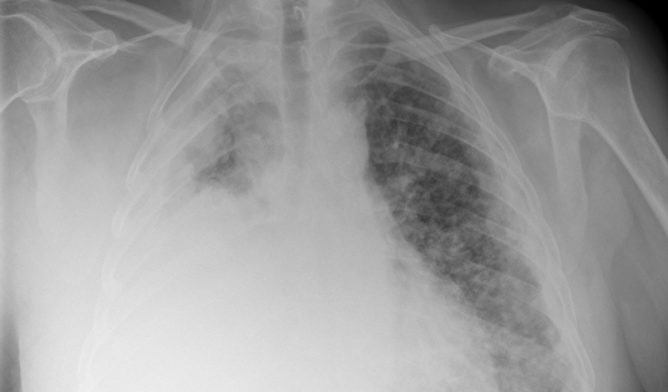
Staging with chest ct, mediastinoscopy, and mri or sometimes with positron emission tomography (pet) and bronchoscopy. These abnormalities may include thickening of the lung lining or fluid buildup in the chest wall. The effusion in the image above was drained. (same patient as image above). Given the presence of the mesothelium in differen. Looks great shape and normal antung. P baas and others on behalf of the esmo. There is no cure for mesothelioma, but treatment can lessen symptoms. Mesothelioma is a rare form of cancer typically affecting the lining of the lungs. Pleural fluid cytology or pleural biopsy. At the screening program at a radiology clinic usa examples of cases in men age 40 with a chest x ray findings mesothelioma. However, the presence of a pleural effusion could also. It's usually linked to asbestos exposure.
Pleural fluid cytology or pleural biopsy mesothelioma x ray. (same patient as image above).

0 comments